The Story of a 'Bright' Concussion Imaging Project

What motivates anyone to do medical research? Sometimes the motivation is simply curiosity. Sometimes you want to have an impact. Sometimes it is because someone you know has a condition that drives you to work on that problem.
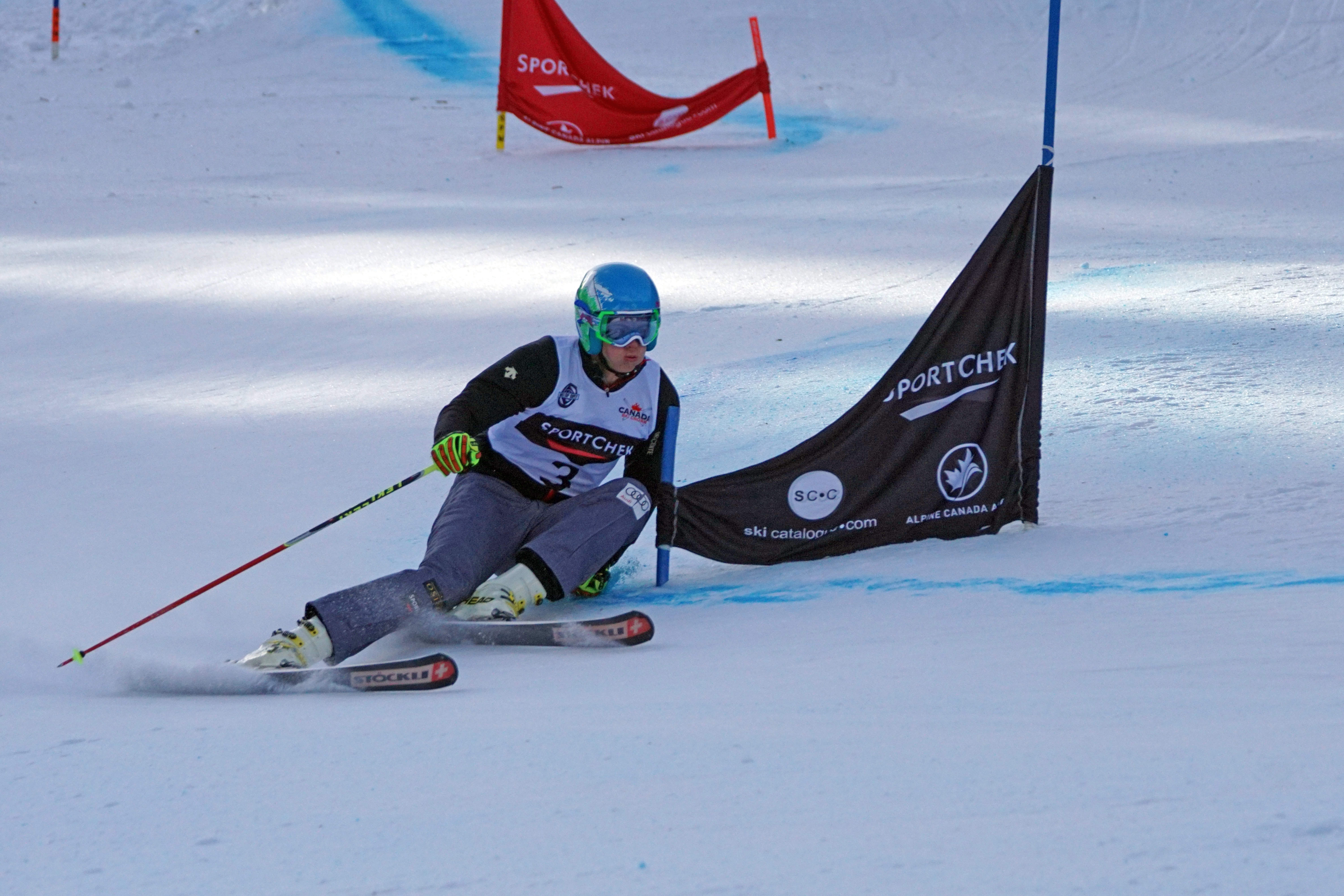
I started work on concussion (also called mild traumatic brain injury or mTBI) when I got involved, as a parent, in ski racing. Young people were getting concussions and it was frustrating, as an imaging scientist, to acknowledge that no current medical imaging method could be used to diagnose or monitor concussion. Surely we could do better! So, I thought about the tools at hand and my experience. Good research draws on the unique skill sets that accumulate over years of doing research. I have a few years under my belt so I should be able to come up with something. Given the low funding levels, and the certainty that I would not be funded to do research on a subject that I had not published on, no matter how good my idea was, I had to think “shoestring”. I had to use the tools already in the lab, convince clinical collaborators to come on board and find staff to do the work (preferably without being paid!). Hmm.
At the time, I was working on applying a new imaging method called functional near-infrared spectroscopy (fNIRS) to study brain activity. I was applying the tool to different problems, largely to develop proof of principle. I had just finished a project with the Multiple Sclerosis group using fNIRS as a marker of reduced brain communication—thinking that this would be a good measure of reduced function that occurs in MS with loss of the sheath around nerves (demyelination), and loss of the nerves themselves (atrophy).

Here are the pieces that I had in place.
- fNIRS detects changes in the amount of hemoglobin (the compound that carries oxygen in the blood) that is bound to oxygen. The form without oxygen (deoxyhemoglobin) has a different color than the form with oxygen (oxyhemoglobin). This is clearly evident when you look at arteries and veins. Arteries are red (high oxyhemoglobin) and veins are bluish (high deoxyhemoglobin).
- As brain activates, oxyhemoglobin goes up and deoxyhemoglobin goes down. As a result, you can use fNIRS to detect areas of brain activity by measuring the changes in amounts of these compounds.
- Regions of brain have a low oscillation of oxyhemoglobin and deoxyhemoglobin in the range of 1 cycle every 2-10s. Regions of brain that are in communication with each other tend to have similar frequencies of oscillation.
- The parts of the brain involved in motor action and sensation are above the ears on both sides of the brain—the motor cortices. If you finger tap with the right hand, the left hand motor cortex activates with an increase in oxyhemoglobin.
- The motor cortices are always talking to each other in an awake healthy person.
- Thus, if you measure the oscillations of oxyhemoglobin and deoxyhemoglobin and compare frequencies in motor cortices, you can obtain a measure of the current level of communication. If you measure a group of control subjects, this value can become a marker of the current state of health. We scaled the value from 0 to 1, and call it coherence (the similarity in frequency between the two cortices).
- We measured a group of MS patients as well as controls. Sure enough, the coherence value was lower in MS patients. I’d like to think that someday this could become a new test for the extent of impairment in MS—but we don’t have funding to test that yet.
So that was the situation as I was pondering how to improve imaging of concussion for these little ski racer dudes and dudettes.
So here is what I was thinking. If shaking the brain damages the linkages between brain regions, then this might be a new method to detect and monitor concussion. I worked out that I could get some good pilot data for less than 50K-the amount needed to hire a MSc student for 2 years, to purchase some supplies and to send the student to one meeting to convey our results. I applied for funding—and got turned down. Hmm. Not abnormal. To all those scientists out there–you are not a bad scientist just because your grant wasn’t funded. Say that again, this time with feeling!
I then got a call which had a lasting impact on my research program. Dr. Jong Rho had read the proposal. He worked at the Alberta Children’s Hospital in Calgary. He let me know that he liked the idea even though it wasn’t funded. He helped me obtain the needed money. Good story eh .
I then went about recruiting and building the team. Dr. Karen Barlow at the Alberta Children’s hospital agreed to help. Karolina Urban, a student from the University of Toronto applied — she had the drive to get the job done. At the time, she was one of Canada’s few professional woman hockey players. She wanted to do the research because of both personal experience and the fact that she knew imaging could have a big impact.
Fast forward a few years. Karolina’s MSc was successful. She showed, in a small group of adolescents with long term concussion symptoms, there was a reduction in coherence measured using fNIRS. In theory, we could scale the hardware to make it portable and write software to make it easy to use. We needed bigger bucks. Armed with the data, and a paper in a good journal (Journal of Neurotrauma), I went after funding to repeat the study, to include adults and to streamline the method. I was successful.
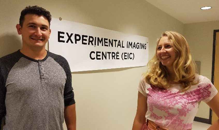
Now Karolina is burning up a storm doing her PhD in concussion research at the Bloorview Hospital in Toronto. I obtained the needed funding to expand to a 3 year program and have recruited another graduate student (Chris Duczynski) and a post-doctoral fellow from Germany (Dr. Lia Hocke).
And we are off—working on a project to develop a portable, safe, sensitive method to monitor brain injury with light. Tune in next year .
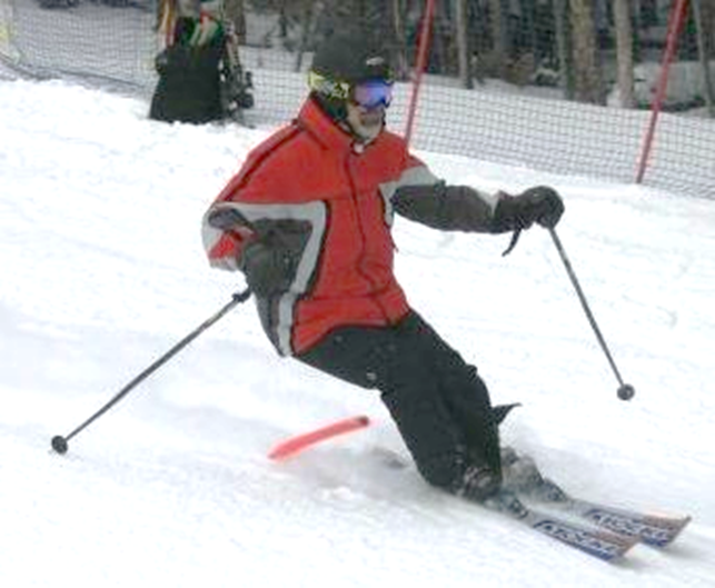
About Dr. Jeffrey Dunn:
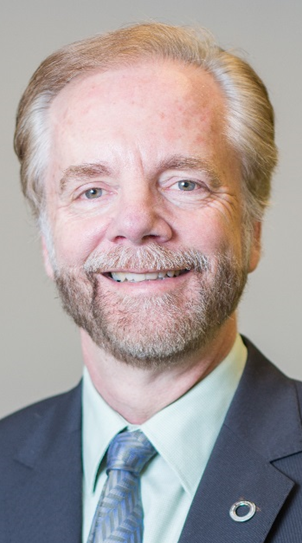
Dr. Jeff Dunn is a Professor in the Department of Radiology, with adjunct positions in the Departments of Physiology and Pharmacology and the Dept. of Clinical Neuroscience. He was recruited from Dartmouth Medical School in 2004. He is a member of graduate programs in Neuroscience, Mountain Medicine and High altitude physiology, Medical Science (including musculoskeletal imaging) and Biomedical Engineering. He works on imaging development (MRI, near-infrared spectroscopy) and focuses on how hypoxia impacts disease progression (Multiple sclerosis, stroke, traumatic brain injury, brain cancer). The lab also undertakes high resolution MR microscopy.
If you would like to learn more about Dr. Jeff Dunn’s research or interested in participating, then please check out the Dunn Lab Imaging page at http://www.ucalgary.ca/dunnimaging/. Or check out Dr. Dunn’s blog at http://jefffdunn.blogspot.ca/.
The opinions expressed in this blog post are the author’s own.